Posts

Showing posts with the label medical imaging

Positron Emission Tomography (PET) Scanners; a Medical Imaging Technique Used to Look For Disease in the Body

Image
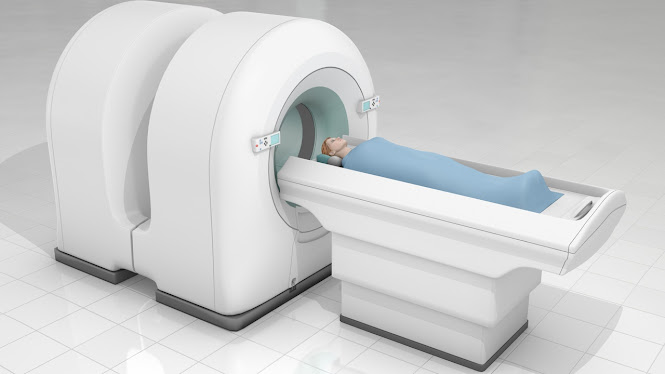
  Positron emission tomography (PET) scanners (an imaging test) help reveal the biochemical or metabolic function of the body organs and tissues. PET scan uses tracer (a radioactive drug) to show normal, as well as abnormal, metabolic activity. Positron emission tomography (PET) scanners are used in the diagnosis of various diseases, such as Alzheimer’s disease and cancer. The basic principle of positron emission tomography involves the detection of a pair of gamma rays emitted indirectly by a radiotracer. PET scanners can also distinguish between malignant tumors and non-malignant tumors. Generally, positron emission tomography (PET) scanners takes about 40 minutes to complete the scan and also measure glucose levels in the body. PET scans provides vital information about tissues and organs, size, shape, and the position. Moreover, PET scanners help detect the abnormal metabolism of tracers in diseases before the disease shows up on other imaging tests, such as magnetic resonance i

A type of radiation therapy, proton therapy is used to treat cancer. It treats cancer using protons rather than x-rays.

Image
  Proton therapy is a kind of radiation therapy — a therapy that utilizes high-energy shafts to treat tumors. Radiation therapy utilizing X-beams has for some time been utilized to treat malignancies and noncancerous (kindhearted) tumors. Proton therapy is a more current kind of radiation therapy that utilizes energy from emphatically charged particles called protons. Proton therapy has shown guarantee in treating a few sorts of malignant growth. Studies have proposed that proton therapy might cause less incidental effects than conventional radiation, since specialists can more readily control where the proton radiates store their energy. In any case, scarcely any investigations have straightforwardly analyzed proton therapy radiation and X-beam radiation, so it's not satisfactory whether proton therapy is more successful in delaying lives. A machine called a synchrotron or cyclotron speeds up protons. The high velocity of the protons makes high energy. This energy makes the p

Chemiluminescence imaging technology has been widely used in high-throughput detection of a variety of analytes due to its excellent sensitivity

Image
  Chemiluminescence is the way toward changing over compound energy into noticeable light that is radiated as the aftereffect of substance (oxidation or hydrolysis) response. This cycle offers delicate and savvy location strategies to a few radio-isotopic and fluorescence procedures and most chromogenic recognition measures. Chemiluminescence imaging is the way toward catching the series of pictures during recognizable proof of low plenitude proteins. Chemiluminescent imaging empowers precise evaluation of Western blotchs with a constant showcase. Chemiluminescent discovery for western smudging is mainstream since it offers a few benefits over other recognition strategies. Utilizing chemiluminescence permits numerous openings to be made, which empowers streamlining of sign to commotion. The recognition reagents can be eliminated and the whole smudge reprobed to imagine another protein or to upgrade discovery of the primary protein. An enormous direct reaction range permits identific

Medical X-Ray Film Processor; the Most Important Part for the X-Ray Image Processing

Image
  There are two types of medical X-ray film processor, such as semi-automatic film processor and fully automatic film processor. These processors are used to decode the information carried by X-ray beam, when they are passed through the tissues. Medical X-ray film processors capture the invisible image into visible form. An automatic device is designed to move X-ray films from one solution to another, without the need for human intervention except to insert a film or cassette. Medical X-ray film processor, whether it is manual or automatic, comprises five basic steps, such as drying, washing, fixing, rinsing or stop bath, and developing. Medical X-ray film processor is the kind of processor that can process images from X-rays, CT scans, MRI's, and it can even process an X-ray film. This kind of development is a lot complicated because it involves a lot of things, including the design of the camera, the software, the data capture and all of this is done with the help of the X-ray

Digital Fluoroscopy System; Used to Examine the Anatomy and Function of Various Body Parts

Image
  Digital fluoroscopy systems are utilized to make digital pictures of the human body. It is type of X-beam used to picture profound constructions of the human body. The system gives itemized pictures of imperative organs like stomach, bladder, heart muscle, and digestive tract. It records a progression of pictures to the PC. Digital fluoroscopy system is broadly used to look at and assess the gastrointestinal parcel, including the colon, duodenum, throat, and the stomach. In addition, digital fluoroscopy is utilized to analyze blockages, aggravation, reflux, scarring, hiatal hernias, ulcers, and tumors. Moreover, digital fluoroscopy system inspects/assesses renal capacity in angiography and venography strategies and agony the executives techniques. Altered barium swallow study, lumbar cut, discography, cardiology, urological medical procedure, situation of weighted taking care of cylinder and incidentally embedded focal catheter, angiography, podiatric medical procedure, and muscula

Dental loupes and cameras are in high demand in Asian countries like Japan, South Korea, and China

Image
  Dental loupes and cameras are utilized to make it simpler to see the internal parts of teeth during dental methodology. They are comprised of a metal casing and different measures of inward focal points that can shine light to give a more prominent perspective on the inside of the mouth. The interior focal points are movable, empowering the dental specialist to change the amplification on a case by case basis without reseting the unit. The demand for dental loupes and cameras is high in Asian nations like Japan, South Korea, and China. This can be credited to high number of dental specialists and expanding center around stylish allure among the general population around there. Dental loupes are ordinarily utilized by orthodontists, yet as of late they have been embraced by restorative dental specialists who utilize the gadget to make more modest models of dentures. Dental loupes are regularly utilized by kids and grown-ups the same, who can see the internal parts of their mouths i

Orthopedic Imaging Rising prevalence of bone-related disorders

Image
  Clinical imaging in orthopedics helps doctors to non-intrusively investigate the body's inward life structures to help in diagnosing an ailment. By delivering pictures reflecting inward anatomical designs of the patient's body, clinical imaging gadgets perceive and track illnesses or wounds. These gadgets are utilized as an information instrument that assists specialists with distinguishing the sickness and the therapy reaction. Orthopedic clinical imaging offers dependable, available, and great imaging, a basic boundary for clinical dynamic for doctors, which might actually decrease the requirement for surgeries. Utilization of orthopedic imaging hardware is exceptionally urgent for diagnosing and treating wounds, infections, and sicknesses identified with bone, ligament, and related bits of the body. Lately, far reaching progressions are happening with regards to plan and usefulness of orthopedic imaging gear. Moreover, interest for orthopedic imaging hardware has dras

Digital Fluoroscopy System; Used to Examine the Anatomy and Function of Various Body Parts

Image
  Digital fluoroscopy systems are utilized to make digital pictures of the human body. It is type of X-beam used to imagine profound constructions of the human body. The system gives itemized pictures of imperative organs like stomach, bladder, cardiovascular muscle, and digestive tract. It records a progression of pictures to the PC. Digital fluoroscopy system is broadly used to look at and assess the gastrointestinal plot, including the colon, duodenum, throat, and the stomach. Also, digital fluoroscopy is utilized to analyze blockages, irritation, reflux, scarring, hiatal hernias, ulcers, and tumors. Besides, digital fluoroscopy system inspects/assesses renal capacity in angiography and venography methods and agony the executives strategies. Adjusted barium swallow study, lumbar cut, discography, cardiology, urological medical procedure, position of weighted taking care of cylinder and incidentally embedded focal catheter, angiography, podiatric medical procedure, and muscular med

Demand for Dental Loupes and Cameras High In Asian Countries Such As Japan, South Korea, and China

Image
  Dental loupes and cameras are used to make it easier to see the insides of teeth during dental procedures. They are made up of a metal frame and various amounts of internal lenses that are able to focus light to provide a greater view of the interior of the mouth. The internal lenses are adjustable, enabling the dentist to change the magnification as needed without having to reset the unit. The demand for dental loupes and cameras is high in Asian countries such as Japan, South Korea, and China. This can be attributed to high number of dentists and increasing focus on aesthetic appeal among the populace in the region. Dental loupes are typically used by orthodontists, but in recent years they have been adopted by cosmetic dentists who use the device to create smaller models of false teeth. Dental loupes are often used by children and adults alike, who can view the insides of their mouths in much greater detail than is possible with traditional instruments. Dental loupes and camer

Digital Fluoroscopy System; Used To Assess the Anatomy and Function of Different Parts of the Body

Image
  Digital fluoroscopy systems are used to make digital images of the human body. It is form of X-ray used to visualize deep structures of the human body. The system provides detailed images of vital organs such as stomach, bladder, cardiac muscle, and intestine. It records a series of images to the computer. Digital fluoroscopy system is widely used to examine and evaluate the gastrointestinal tract, including the colon, duodenum, esophagus, and the stomach. Moreover, digital fluoroscopy is used to diagnose blockages, inflammation, reflux, scarring, hiatal hernias, ulcers, and tumors. Furthermore, digital fluoroscopy system examines/evaluates renal function in angiography and venography procedures and pain management procedures. Modified barium swallow study, lumbar puncture, discography, cardiology, urological surgery, placement of weighted feeding tube and peripherally inserted central catheter, angiography, podiatric surgery, and orthopedic surgery are some of the some of the proc

Digital fluoroscopy system gaining popularity across screening of common eye disease

Image
  Digital fluoroscopy system is used in a common treatment procedure of eye doctors nowadays. Fluoroscopy as it is commonly called is the process of using x rays of laser to diagnose and observe problems of your ocular tissue. This procedure is generally done for the treatment of newborns but is also useful for diagnosing many eye disorders in older people. This is why a digital fluoroscopy system is beneficial in minimizing the unnecessary exposure of patients to radiation. With the help of laser, doctors can create three-dimensional images of the retina. The advantage of the digital fluoroscopy system lies in its quick availability of x-ray laser and the fact that there is no unnecessary exposure to radiation. Using digital imaging for various health issues has already been around for more than 40 years. In fact, with the help of computed radiography, it allows obtaining better and higher quality images than what would be able to obtain with traditional techniques such as computed