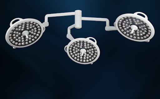
Medical Illumination Systems are used in surgical procedures to shed light on deep surfaces and differentiate the real color of tissues in the cavity
Medical illumination systems offer good visibility on
very deep, narrow, and flat surfaces inside a surgical cavity. Central luminance
of medical illumination systems range from 40,000 to 160,000 lumens. The light
of medical illumination helps to distinguish the actual color of the tissue
within a cavity, due to multiple light points fixed on the same system to
assist in reducing shadowing due to light refraction at different angles. This
method of illuminating the skin enables a bright, detailed image to be made of
the inner portion of the organ or tissue. For this reason, medical illumination
systems are known to be extremely advantageous in surgical procedures where
detailed images of the internal organs and tissues are needed for effective
treatment. For instance, in September 2021, a major manufacturer of laser
systems in the U.S., Leonardo Electronics, Inc., received an automotive IATF
16949 certification for its VCSEL (vertical cavity surface emitting laser) in
medical applications.
Medical
illumination systems come
in two forms: with or without LED lights. In the former, the illuminator units
are permanently fixed into the frame; thus, they are not mobile and are
therefore more effective than portable illuminators. These illuminators use
energy-efficient bulbs that also consume less power than incandescent bulbs. In
addition, medical illumination systems with LEDs produce brighter light than
incandescent bulbs, contributing to faster image resolution.
Medical illumination systems have two basic components: the probe, which
holds the light source; and the illumination source, which directs the beam to
the target. Each of these components is capable of providing different levels
of illumination. The two basic types of medical illumination systems are:
central (one fixed probe) and peripheral (two or more fixed probes). A central
medical illumination system can provide high levels of light for the entire
surgical cavity; whereas, peripheral systems can be placed at various points
around the operating table for targeting specific areas of concern. Regardless
of the type of system used, the important parameters to consider include beam
distance, exposure time, and fluency of the images.


Comments
Post a Comment