Life Cell Imaging is the research of living tissues with a time-lapse microscope to obtain high-resolution of tissue structures
Life cell imaging is widely used by biochemists to gain better
understanding of physiological function through the examination of complex
cellular dynamics. Life cell imaging was first pioneered in the first year of
the new millennium. Life cell imaging has many applications in science,
especially in cell evolution and physiology. In particular, it can be used for
studying cancer cells under living conditions.
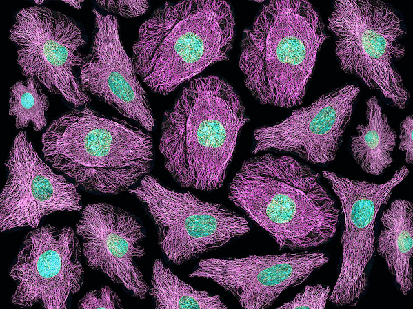
Different techniques are used in the field of live cell
imaging. These include electro-photometry, optical microscopy, confocal
microscopy, chemotaxle and bid analysis, imaging energy, fluorescence
microscopy, cryoepiezo imaging, chemotaxillary tomography, non-disposable
microscopes and confocal microscopes. The confocal microscopes are preferred
due to their clarity, high image quality and fast acquisition time. The
non-disposable microscopes are used in the field of life
cell imaging to observe dynamic processes without affecting the sample
or environment. For instance, in September 2021, a major medical imaging
solutions providing company, NorthStar Medical Radioisotopes, LLC, partnered
with Point Biopharma Global for developing a high-energy emitting radioisotope,
Ac-225.
In the field of cell imaging, two different methods are commonly used to
observe living tissues. One technique consists of using fluorescent proteins as
a marker for the intensity changes of cells. Macromolecules of fluorescent
proteins are detected by the electrons of amino acids that bind them. The other
technique uses the concept of peroxisomes, which are small pores on the cell
membranes.
The imaging systems are designed to observe single macromolecules or
many molecules at a time. There are three types of single marker microscopes
for single molecules, multicellular structures and whole organisms. There are
also high resolution and high volume microscopes available in the life cell
imaging sector. Single molecule image systems are made up of multiple molecules
of fluorescent proteins bound to an electron microscope slide. The techniques
used in single molecule imaging systems include scanning, fluorescence
microscopy and chemical fluorescent labeling.
Multicellular systems include multiple sets of cells
from diverse environments, such as human blood stages. It is important to
observe dynamic processes between cells. There are different systems that can
be used to observe cell biology in multicellular systems. These include
electrophoresis, chemiluminescence and confocal microscopy.


Comments
Post a Comment